
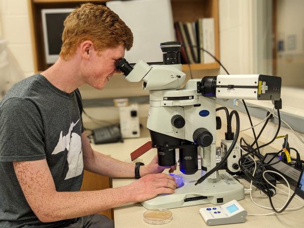
A microscope acquired in spring 2023 using grant funds is opening new opportunities for research and teaching in the Kalamazoo College biology department.
The fluorescence dissecting microscope boasts several advantages over other microscopes in the department, said Michael Wollenberg, associate professor of biology and department chair, and Amanda Wollenberg, associate professor of biology. Its features include optics that allow clear views of individual cells, fluorescence to help differentiate between types of cells, space to manipulate a sample while viewing it, and a dedicated camera and software program.
“We want to be able to look at single bacterial cells, and they’re one-millionth of a meter large, so they’re not visible to the naked eye,” Michael said. “What’s really important is that we have a microscope that has extremely good optics, and those optics resolve the sample very well—they magnify it to the point where we can see individual bacterial cells. Bacterial cells are clear and transparent, and having a set of fluorescent molecules inside the cell allows us to tag them with a glowing marker so we can say, ‘OK, that transparent cell that’s really tiny—that’s actually the bacterial cell we’re interested in, as opposed to schmutz or a eukaryotic cell or some other bacterial cell, and it’s located in X, Y or Z place, which gives us a three-dimensional resolution of the relationship that we’re looking at.’”
While many microscopes use white light to illuminate samples, a fluorescence microscope can do more.
“Bacteria might be too small to actually see just using white light,” Amanda said. “If you genetically manipulate those bacteria so that they glow fluorescent—and that’s a very common technique; people do it all the time, Michael can do it in his lab—if you have a microscope that can detect fluorescence, you can track those bacteria. You can see where they are because they’re glowing green.”
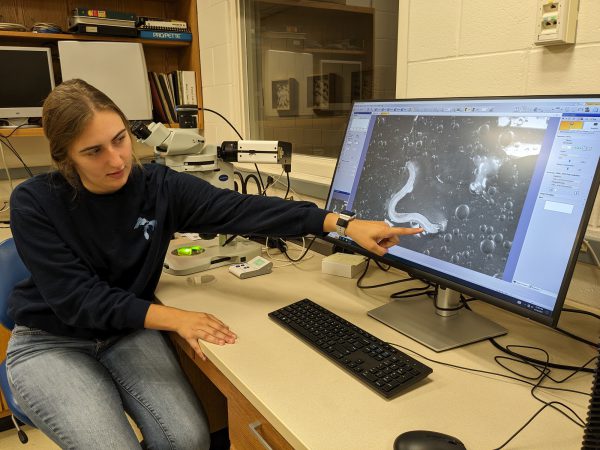
The microscope also has a camera and software program that allows the user to take and analyze photos through the microscope as well as project to a computer screen—two key advantages in research and in teaching that are new to the biology department in terms of dissecting microscopes.
“The camera and software are really important when it comes to trying to share the results of what we see with other scientists by publishing,” Amanda said. “You can’t just say, ‘Oh, trust us, we saw it.’ You must have a picture, and it’s got to be a high-quality picture. Ideally, you also try to quantify some of the information in that picture. That means you don’t just show a picture. You say, ‘We took 100 pictures, and we did this analysis of the intensity of the color, using the software program, and in 70 percent of the pictures, the intensity was higher than a certain threshold.’”
The computer projection makes the microscope an excellent teaching tool.
“A microscope has two eyepieces,” Michael said. “You look in the eyepieces, and you’re the only person who can see what you’re seeing. This camera is a great way to project what the microscope sees to a larger group of people, be it a small research laboratory, like our summer research with undergraduates, or a small class.”
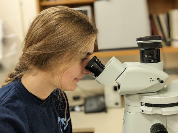
Biology major Allison Sokacz ’24 has worked in Michael Wollenberg’s lab for three summers. She recently started using the microscope in the summer research that will form the basis for her Senior Integrated Project.
“The intensity that lets you see the entire fluorescence from this microscope, versus some of the other scopes we have, is really helpful,” Sokacz said. “It’s a lot easier to see what you have.”
The camera is essential to her project, as she is working with two different forms of a bacteria and will be able to compare their locations using saved images. She also appreciates the benefits of the screen projection.
“Microscopy is hard, because only one person can see,” Sokacz said. “I just took microbiology with Dr. [Michael] Wollenberg this spring, and I really struggled with microscopy, because it’s different for everyone. My lab partner might say, put the zoom to this, but then I might not be able to see it. With this microscope, I can say what I see, and they can also see it, instead of, ‘Well, I saw this but then it moved off the screen,’ or, ‘I can’t get it in focus.’ Being able to show a whole room what you’re seeing is definitely helpful.”
In addition, the microscope has a wheel that allows for different filters that can detect different colors of fluorescence, which expands the future possibilities for use.
“You could label one type of bacteria with green and one with red, and now you can look at the dynamics,” Amanda said. “Are the green ones in one place, are the red ones in another place? Another thing researchers will do sometimes is to stain the host with one color and the bacteria with a different color, and that can help resolve some of the questions of what you are seeing.”

The microscope was purchased using funds from a $400,000 National Science Foundation grant, awarded to the Wollenbergs in June 2018 to study mechanisms of specificity and tolerance in a nematode-bacterial symbiosis. About 9 percent of that budget was for the microscope, which cost about $35,000. The rest of the total includes about 12.5 percent for other materials and small equipment, 5 percent for travel, 26 percent for indirect costs like building infrastructure, and slightly less than half for student pay, summer salary and benefits.
“Science uses very condensed writing, where each word means so much,” Amanda said. “In the title of our research, ‘mechanisms’ tell you that we’re looking at the molecular side of things: not just that this happens, but how does it happen? ‘Specificity’ is getting at this idea that when two organisms are trying to have a relationship with each other, it’s not like just any bacteria can come in and live with any animal. ‘Tolerance’ is, even once they’ve found each other and formed that partnership, they have to keep getting along with each other. From the animal perspective, it can’t start killing off that bacteria like it does to other, more pathogenic bacteria. The specific relationship we’re looking at is a nematode-bacterial symbiosis. This is telling us that the animal is the nematode–that’s a roundworm that lives in the soil–and the partner it has is bacteria. Symbiosis is saying they’re in partnership, they both benefit each other. We’re trying to understand how they find each other and get along with each other within that system.”
The duo is well-suited to the research, with Michael bringing a microbiology perspective while Amanda has an immunology focus.
“The big picture is that we live in a microbial world where there’s lots and lots of microorganisms that are in and on our bodies and all animal bodies,” Michael said. “Basically, they facilitate everything that animals do; we can’t survive without them. Understanding how animals tolerate beneficial microorganisms is a big open scientific question. How do we train our immune systems, or how are our immune systems calibrated, so that the friendly bacteria get into association with organisms and maintain those associations that are beneficial?”
That’s not very well understood, Michael notes, “especially not in really complicated organisms like humans, where there are myriad different species of microorganisms that are associated with us. If you think about our gut, it’s like its own ecosystem. We do research with simple models to try to understand detailed answers to bigger biology questions, in the hopes that other scientists can apply that research to things that are more relevant for human health or other animals that have more complicated associations in their health.”
The new microscope is an invaluable tool in the Wollenbergs’ research, and they also look forward to the whole biology department finding ways to use it in the classroom.
“That was part of the reason we wanted to get this piece of equipment as well, is to integrate it with teaching and use it as a teaching tool,” Michael said. “As we’re coming online with the things we’ve wanted to do with the grant, it’s giving us ideas of how we can translate this into the classroom.”